EMT Practice Test

1. Question Content...


Question List

Question1: A practitioner obtains this image of a metacarpophalangeal joint of a patient with periodic joint swelling and warmth. Which of the following is the MOST likely diagnosis?

Question2: In this volar transverse image of the carpal tunnel, the arrows represent needles, and the arrowheads represent the needle tips. In planning a carpal tunnel corticosteroid injection with ultrasound guidance, which of the arrows combines the safest path through tissues with the BEST needle visualization?

Question3: A 62-year-old man presents with left shoulder pain. The ultrasound image was taken at the patient's maximum abduction (45 degrees). Which of the following rotator cuff pathologies is BEST evaluated with this shoulder position?

Question4: An 81-year-old male with known shoulder osteoarthritis presents to the office for an ultrasound-guided injection of the glenohumeral joint. Prior to injection, where should the needle tip be visualized?

Question5: What is the MAXIMUM normal dimension of the structure indicated by the line in this image?

Question6: A 41-year-old man presents with suspected right carpal tunnel syndrome but normal nerve conduction studies. For which reason will a sonographic study be helpful for further diagnostic evaluation?

Question7: Which of the following is the MOST appropriate next step to confirm the presence of pathology in this metacarpophalangeal joint?

Question8: At approximately how many degrees of flexion is the MOST sensitive resting position of the knee for imaging fluid in the suprapatellar recess?

Question9: A practitioner is counseling a patient with suspected rotator cuff tendinopathy on imaging options. Which of the following is CORRECT regarding diagnostic ultrasound examination?

Question10: In this image of a knee, what is the MOST obvious abnormality?

Question11: For which of the following conditions is the use of diagnostic musculoskeletal ultrasound appropriate?

Question12: What should be taken into consideration prior to any procedure?

Question13: Which of the following statements regarding ultrasound probe care is MOST accurate?

Question14: In this ultrasound image of a hip, where is the transducer positioned?

Question15: In performing an in-plane ultrasound-guided procedure, it is helpful to orient the transducer so that the:

Question16: This suprapatellar longitudinal scan in a 3-year-old girl is MOST consistent with which of the following?

Question17: Which carpal bone is visualized medially to the ulnar nerve on a transverse scan of the wrist at the level of the distal wrist crease?

Question18: How reliable is ultrasound as compared to other imaging modalities for diagnosing synovitis in the joints of the fingers?

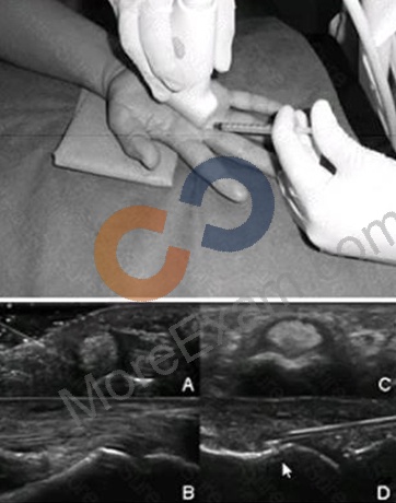
Question19: A photograph of an ultrasound-guided injection approach is shown below. With which ultrasound image does it BEST correlate?

Question20: Why is musculoskeletal ultrasound (MSKUS) useful in monitoring the treatment response in rheumatoid arthritis?

Question21: The orthogonal images of a supraspinatus tendon shown here are MOST consistent with which of the following?

Question22: What is the position of the arm MOST likely used to obtain this B-Mode image of the shoulder?

Question23: Which of the following images is MOST consistent with crystal-induced arthropathy?

Question24: How is the dynamic evaluation for subacromial impingement and adhesive capsulitis performed?

Question25: Which of the following is MOST likely to cause vertical lines on every image obtained from a piece of ultrasound equipment?

Question26: Which of the dorsal longitudinal metacarpophalangeal images below MOST suggests the diagnosis of rheumatoid arthritis?

Question27: A 38-year-old woman with recent history of right shoulder subacromial impingement presents for a two-week follow-up after an anatomically guided subacromial/subdeltoid corticosteroid injection. She reports some initial, but only short-lived, improvement of her right shoulder pain and discomfort with arm elevation.
Her right shoulder X-rays are normal. What would be the MOST appropriate next intervention?

Question28: Which probe is the BEST choice for ultrasound examination of the adult wrist?

Question29: A 30-year-old pregnant woman with rheumatoid arthritis presents with right wrist pain and swelling despite a palpation-guided corticosteroid injection one month ago.

Question30: Which of the following anatomic structures BEST fits the scope of rheumatologic ultrasound practice?

Question31: Which of the following is shown in this longitudinal image of the talonavicular-cuneiform joints in a
15-year-old girl?

Question32: A 72-year-old male with morbid obesity and a known history of knee osteoarthritis presents for an ultrasound-guided knee injection. From which of the following sites should the needle be inserted so that the needle path can be BEST visualized?

Question33: Which effect is achieved by increasing the gain while in Doppler mode?

Question34: The image below shows the injection of the dorsal wrist region in a patient with rheumatoid arthritis. The needle is indicated by the arrows. Considering the ultrasound findings, which of the following is MOST appropriate in completing the injection?

Question35: A 30-year-old female patient with rheumatoid arthritis reports pain at the medial malleolus. The medial side of the ankle is tender and seems to be swollen. How should a practitioner interpret the transverse images at the medial malleolus?

Question36: A patient with diabetes presents with right ankle pain and swelling. The practitioner is unsure of the cause and decides to perform a musculoskeletal ultrasound examination. Which of the following in the practitioner's report is the BEST statement of indication for this study?

Question37: Which statement is TRUE regarding the techniques that can minimize infection during ultrasound-guided procedures?

Question38: Which is the MAIN pathologic finding shown by this ultrasound picture in a patient with psoriatic arthritis?

Question39: A 78-year-old woman presents with three days of ankle pain and swelling. Her exam reveals swelling localized around the medial malleolus, and any motion or palpation causes pain. The medial transverse ultrasound view of the ankle is shown. Based on her presentation and the ultrasound image, what is the MOST likely anatomic structure from which her pain is emanating?

Question40: What should be adjusted to improve the quality of the image of the tibiotalar joint?

Question41: The best way to facilitate visualization of ultrasound images in an exam room.

Question42: Which of these supraspinatus longitudinal images has the BEST combination of ultrasound machine settings?

Question43: By comparing gray-scale images from before pharmacologic treatment and after months of treatment, rheumatologists are able to assess patient improvement and response with regard to: